| Abstract |
The hereditary condition known as ectodermal dysplasia is characterized
by the absence or defect of two or more ectodermally derived structures. The
most commonly observed forms of ectodermal dysplasia are the hidrotic and
hypohidrotic types; discrimination is based on the absence or presence of sweat
glands. A case of 8-year-old male child with hypohidrotic ectodermal dysplasia
with complete anodontia of primary as well as secondary dentitions is
presented. The child had a short stature, low intelligent quotient (I.Q.,), and
was underweight. The patient experienced episodes of high fever, was intolerant
to heat, and did not sweat. He exhibited smooth and dry skin, sparse
light-colored eyebrows. Dental clinicians can be the first to diagnose
ectodermal dysplasia due to the absence of teeth.
Introduction |
Ectodermal dysplasia is a rare congenital hereditary entity.The ectodermal dysplasia represents a group of inherited conditions in which two or more ectodermally derived anatomic structures fail to develop. Ectodermal dysplasias' represent a large and complex group of diseases comprising of more than 170 clinical conditions.
Depending upon the presence or absence of sweat glands, it is divided into the hidrotic (Clouston syndrome) and anhidrotic types. Latter variety is characterized by anhidrosis, hypodontia, and hypotrichosis.(CST-syndrome i.e. Christ- Siemen-Touraine syndrome and anhidrotic/hypohidrotic, ectodermal dysplasia being synonymous.)
In most cases, this disorder seems to show an X-linked pattern, with gene mapping to Xq12-q13.1; therefore, a male predominance is usually seen. Female patients may show partial expression of this abnormal gene. Individuals affected by it show the triad comprising anhydrosis/hypohydrosis, hypotrichosis, and dental hypoplasia. The patients present a soft, smooth, thin dry skin.
Hypodontia has been considered to be a multifactorial condition with genetic and environmental influences, and published opinions differ on the importance of each factor. Larmour et al., state that recent developments in molecular genetics are revealing the roles of the homeobox genes in the control of the complex epithelial/mesenchymal interactions that occur during dental development. Those of particular interest for dental development are the muscle-specific homeobox genes, Msx1 and Msx2.
Case Report
|
Here is the case report of a 8-year-old male child with ectodermal dysplasia. The chief complaint of the patient was difficulty in mastication due to absence of maxillary and mandibular teeth. His personal history revealed that teeth never erupted. His mother told that his birth was normal and family history was negative. The patient experienced episodes of high fever, was intolerant to heat, and did not sweat. He was unable to directly look towards a light source. Physically, the child had a short stature and was underweight. His I.Q., level was low, and it was evident when asked about schooling. The child had sparse light-colored eyebrows(Figure 1). Nails were thin and brittle (Figure 2) and (Figure 3). Skin was dry and rough (Figure 1), (Figure 2) and (Figure 3). Other characteristics of ectodermal dysplasia, such as frontal bossing, saddle nose, reduced vertical dimension of face due to total anodontia were also noticed.
Intraoral
examination revealed absence of teeth with thin alveolar crests (Figure 4) and (Figure
5). Oral mucosa appeared
dry; otherwise, no mucosal defects were seen. The tongue and palate appeared
normal.

Figure 1:
Child showing dry skin, sparse eyebrows, eyelashes, and scalp hair

Figure 2: Hands showing dystrophic (thin and brittle)
nails
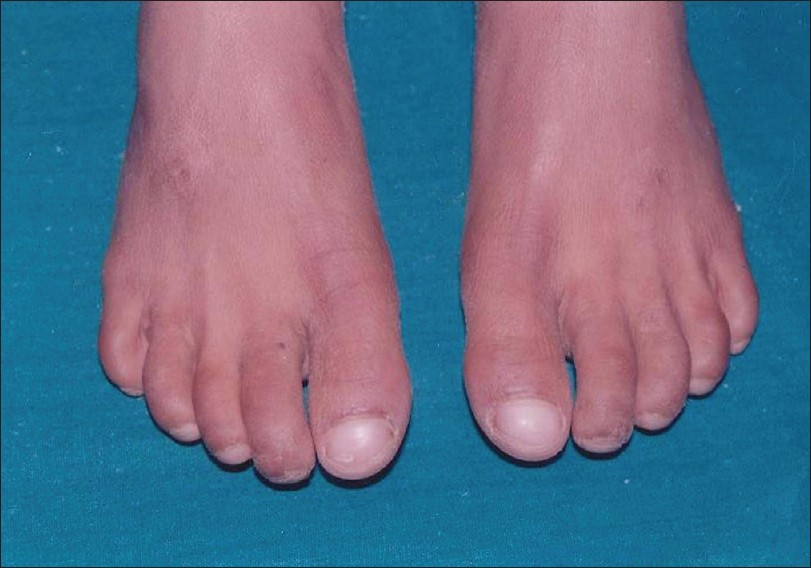
Figure 3: Feet showing dry skin and dystrophic nails

Figure 4: Complete anodontia of maxillary arch

Figure 5: Complete anodontia of mandibular arch
Occlusal and panoramic radiographs revealed no primary and
permanent teeth (Figure 6), (Figure 7) and (Figure 8). In
order to improve mastication and aesthetics, both upper and lower complete
dentures were fabricated (Figure
9).

Figure 6: Occlusal view of maxilla

Figure 7: Occlusal view of mandible

Figure 8: Orthopantomogram (OPG) showing complete anodontia

Figure 9: Post treatment photograph
Discussion
|
Hypohidrotic
ectodermal dysplasia is characterized by hypohidrosis, hypotrichosis, and
hypodontia. Ectodermal dysplasia is one of the most important
anomalies of interest to dental clinicians because of the absent or misshapen
teeth.
Hypodontia is known as one of the major factors of ectodermal dysplasia and is almost always present. In severe cases, no teeth form. The absence of primary teeth (true anodontia) is a rare phenomenon. In this case, the patient's history and clinical and radiographic examination revealed the absence of primary teeth. Acikgoz et al., and Vieira et al., also reported true anodontia of primary teeth. Total anodontia denoted by complete developmental absence of teeth in both primary and secondary dentitions was reported Pirgon et al., and Pannu and Singh. Radiographs showed no signs of formation of tooth buds. It is claimed that primary teeth must be present for the development of their permanent successors. There are no permanent teeth in the oral cavity of the patient, similar finding reported by Vieiraet al.
The patient experienced episodes of high fever, was intolerant to heat, and did not sweat. The patient had sparse eyebrows. Child's inability to perspire, more comfortable during cold weather, and absence of hair from the eyebrows with scanty eyelashes were also reported by Gupta et al., and Pannu and Singh (2002).
Short stature, underweight in relation to age and mental retardation were reported in accordance to case reported by Gupta.
Management: To improve the appearance, mastication, and speech, the child was provided with maxillary and mandibular complete dentures similar to treatment provided by Vierra et al., Pannu and Singh. The dental team should be aware of the clinical presentation of ectodermal dysplasia in order to provide the correct guidance for functional, social, and psychological needs of the patients.
References
|
1.
|
Acikgoz A, Kademoglu O, Elekdag-Turk S, Karagoz F. Hypohidrotic
ectodermal dysplasia with true anodontia of the primary dentition.
Quintessence Int 2007;28:17-23.
|
2.
|
Neville BW, Damm DD, Allen CM, Bouquot JE. Dermatologic diseases. Oral
and Maxillofacial pathology, 3 rd ed. Noida: Elsevier
Publications; 2009. p. 741-3.
|
3.
|
Yavuz I, Kiralp S, Baskan Z. Hypohidrotic ectodermal dysplasia: A case
report. Quintessence Int 2008;39:81-6.
|
4.
|
Suprabha BS. Hereditary ectodermal dysplasia: A case report. J Indian
Soc Pedo Prev Dent2002;20:37-40.
|
5.
|
Gupta S. Christ-siemen-touraine syndrome-A case report. Journal of
Indian dental association2003;74:533-7.
|
6.
|
Rajendran R. Diseases of the skin. In: Shafer WG, Hine MK, Levy BM,
Rajendran Rand Sivapathasundharam editors. A text book of oral pathology. 5 th ed.
New Delhi: Elsevier Publications;2006.p.1099-102.
|
7.
|
Larmour CJ, Mossey PA, Thind BS, Forgie AH, Stirrups DR. Hypodontia-A
retrospective review of prevalence and etiology, Part 1. Quintessence Int
2005;36:263-70.
|
8.
|
Vieira KA, Teixeira MS, Guirado CG, Gaviao MB. Prosthodontic treatment
of hypohidrotic ectodermal dysplasia with complete anodontia: Case report.
Quintessence Int 2007;38:75-80.
|
9.
|
Pirgon O, Atabek ME, Tanju IA. Congenital anodontia in ectodermal
dysplasia. J Pediatr Endocrinol Metab 2008;21:1111-2.
|
10.
|
Pannu K, Singh BD. Ectodermal dysplasia with total anodontia:
Rehabilitation of a seven year old child. J Indian Soc Pedo Prev Dent
2002;20:114-7.
|
11.
|
Gupta HL, Singh H, Prabhakar BR. Anhidrotic hereditary ectodermal
dysplasia. Indian J Pediat 1965;32:173-5.
|
No comments:
Post a Comment